
Transmission Electron Micrograph of a White Blood Cell

Ultrathin section through a white blood cell. Transmission Electron Microscope, 1000:1 (when 15cms wide)
Scanning Electron Micrograph of Bone

Surface of a bone with osteoporosis (beige). Scanning Electron Microscope, 1000:1 (when 15cms wide)
SEM image of the Degistive Tract

Surface of the mucous membrane of the stomach (gaster) . Scanning Electron Microscope, 230:1 (when 15cms wide)
Freeze Fraction of Fatty Cells

Fat cells (adipocytes) surrounded by connective tissue. Scanning Electron Microscope, 600:1 (when 15cms wide)
Freeze-Fraction of a Tooth seen with the SEM

The inner cave of a tooth is called pulpa. It is full of odontoblasts and blood vessels that care for the tooth. Freeze flaction, Scanning Electron Microscope, 7000:1 (when 15cms wide). Prepublication “Visual Biology in Dental Medicine” by Quintessence Publishing
SEM image of single Enamel Cells.
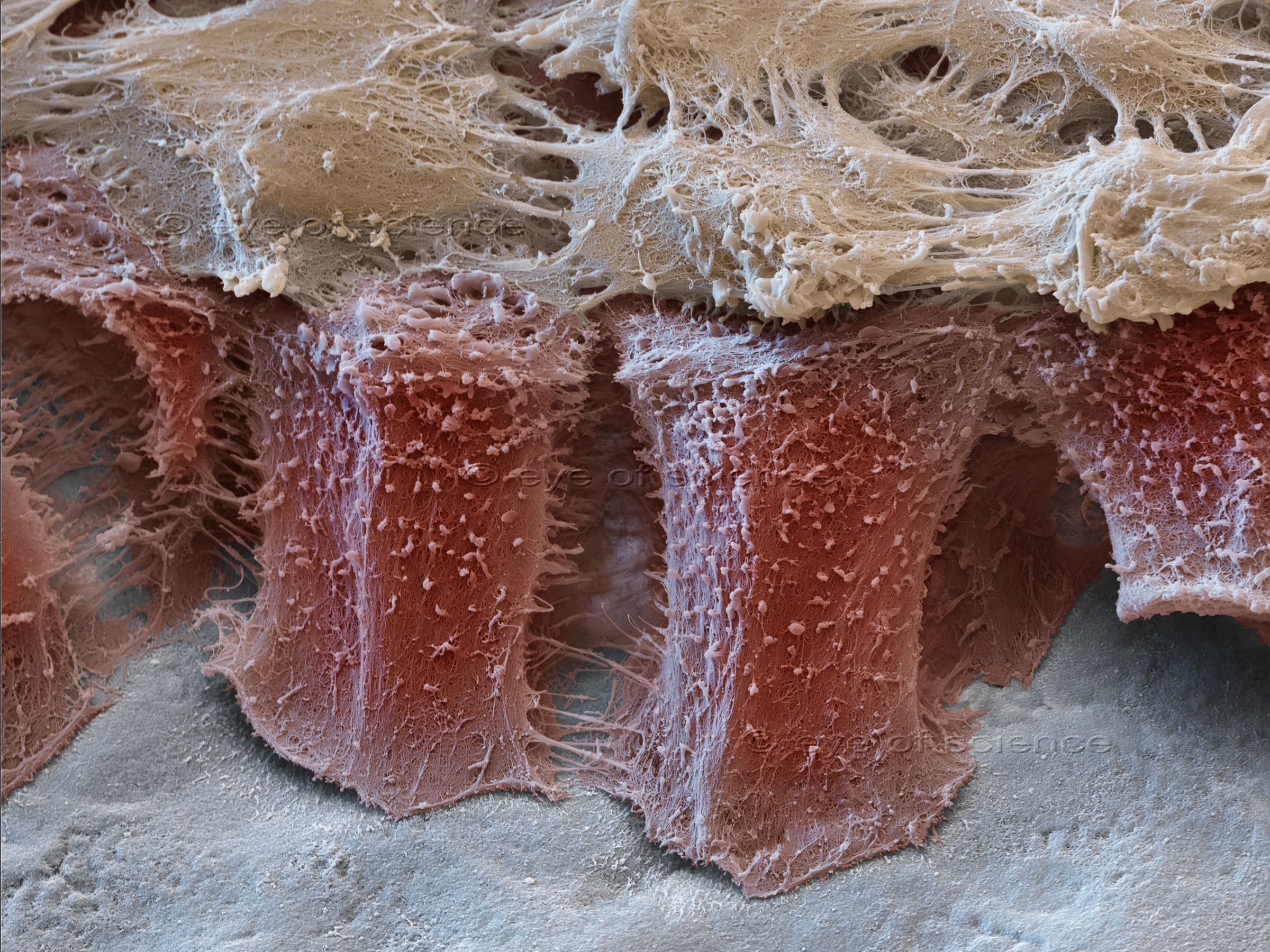
Ameloblasts (red) on the developing tooth create the tooth enamel. Scanning Electron Microscope, 3500:1 (when 15cms wide). Prepublication “Visual Biology in Dental Medicine” by Quintessence Publishing
Scanning Electron Microscope image of a Hair
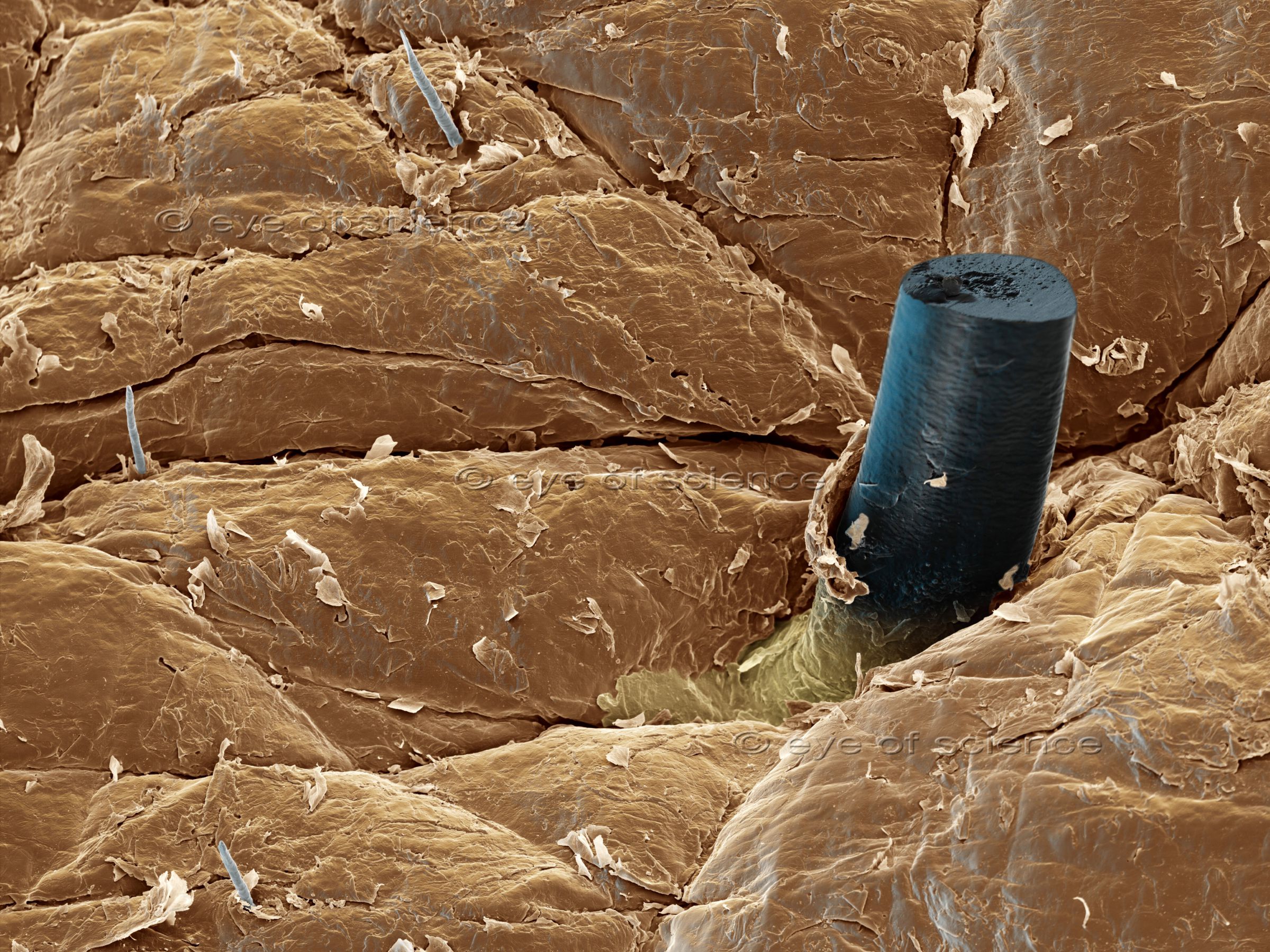
Skin surface with a beard hair that has been cut one day before. Scanning Electron Microscope, 120:1 (when 15cms wide)
Scanning Electron Micrograph of Blood

Red blood cells and one Lymphozyte. Scanning Electron Microscope, 5000:1 (when 15cms wide)
Coloured SEM image of a Bronchus

Section of a bronchus with cilia (yellow) on top of the mucous membrane cells (red) and connective tissue (grey) containing two blood vessels. Scanning Electron Microscope, 1250:1 (when 15cms wide)
Microscopic View into a Lung

View into a lung alveolus. In its depth capillary blood vessels are visible. Scanning Electron Microscope, 250:1 (when 15cms wide)
SEM image of Lung Surface

Surface of a lung alveolus. The capillary blood cells can be seen through the extremely thin walls of the capillaries. Scanning Electron Microscope, 1800:1 (when 15cms wide)
Cancer Therapy, Cells seen with the SEM.

A cancer cell is attacked by a chimeric antigene receptor cell (CAR cell). Scanning Electron Microscope, 3600:1 (when 15cms wide)
Freeze Fraction of Plaque, seen with an Electron Microscope
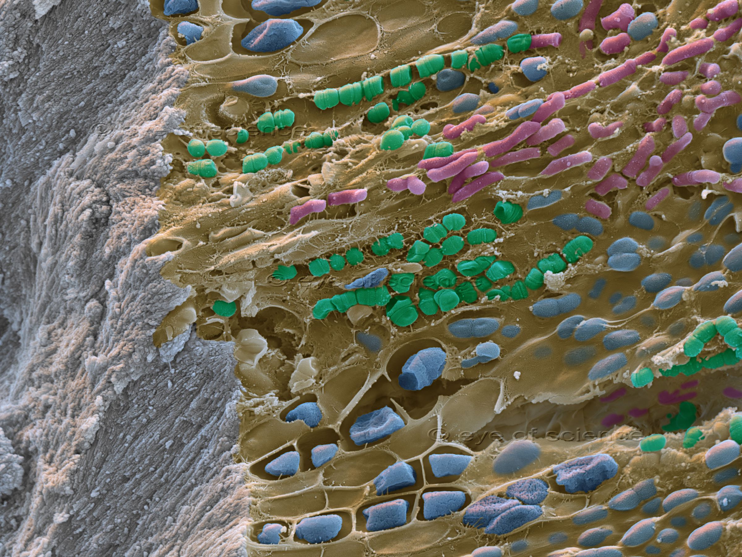
Cross section of a tooth (left, grey) with plaque. The tooth is covered with a thick biofilm of bacteria. Scanning Electron Microscope, 7000:1 (when 15cms wide)





© Copyright for all images on this site by eye of science. All rights reserved.