
Plants and Soil seen with Scanning Electron Microscope
The scanning electron microscope opens up worlds hidden in the smallest of our environment. With microphotography we identify rust fungus, spores, trichomes or visualize the distribution of active substances on plant surfaces. Crop science industry benefits from the depiction of microscopic details of plants, soil components or agents.
Das Rasterelektronenmikroskop eröffnet Welten, die im Mikrokosmos unserer Umwelt verborgen sind. Mit Mikrofotografie dokumentieren wir Rostpilze, Sporen, Trichome oder visualisieren die Verteilung von Wirkstoffen auf Pflanzenoberflächen. Die Agrarindustrie profitiert von der Darstellung mikroskopischer Details von Pflanzen, Bodenbestandteilen oder Wirkstoffen.
Micro Capsule
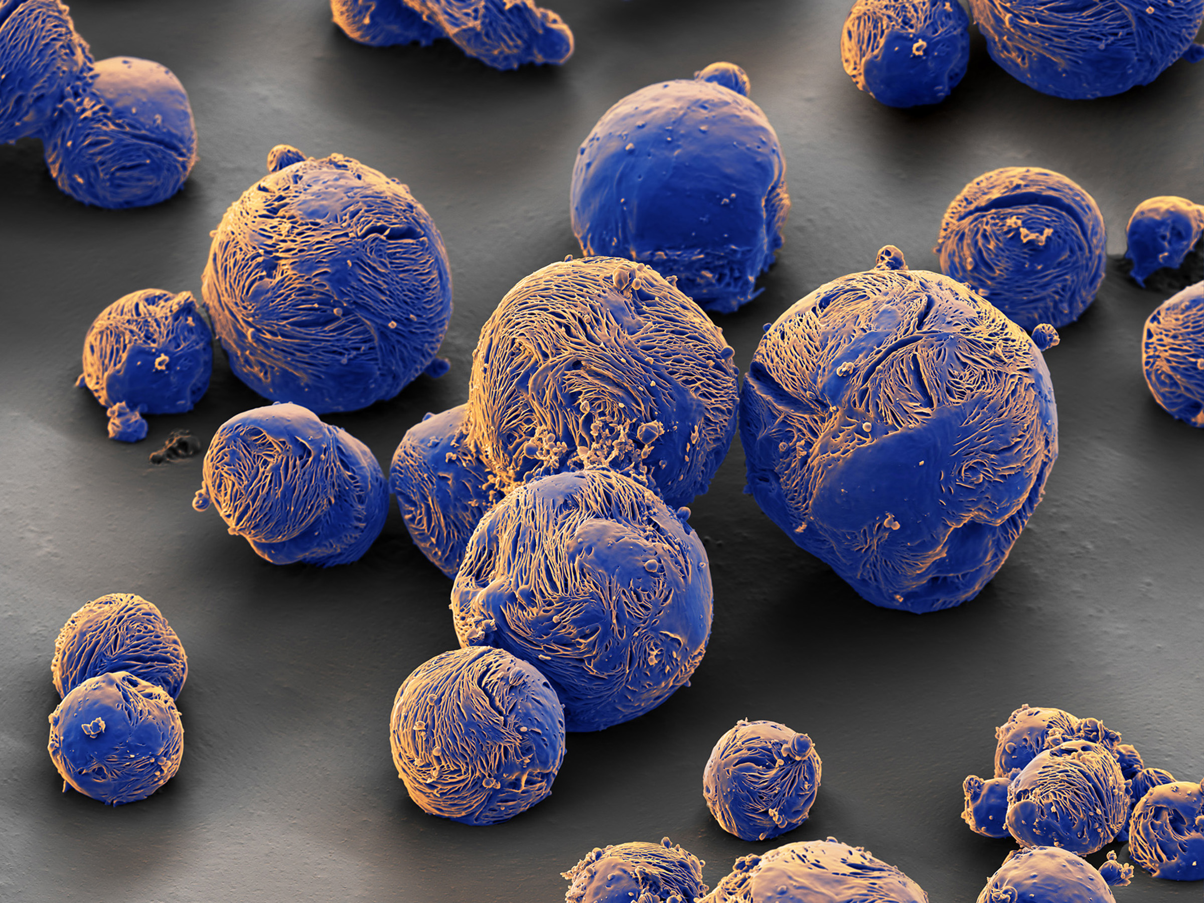
SEM image of a micro-capsule made of polycaprolactam. These capsules were developed to delay the release of pesticides at the site of action.
REM-Aufnahme einer Mikrokapsel aus Polycaprolactam. Diese Kapseln wurden entwickelt, um die Freisetzung von Pestiziden am Wirkungsort zu verzögern.
Rust Fungus
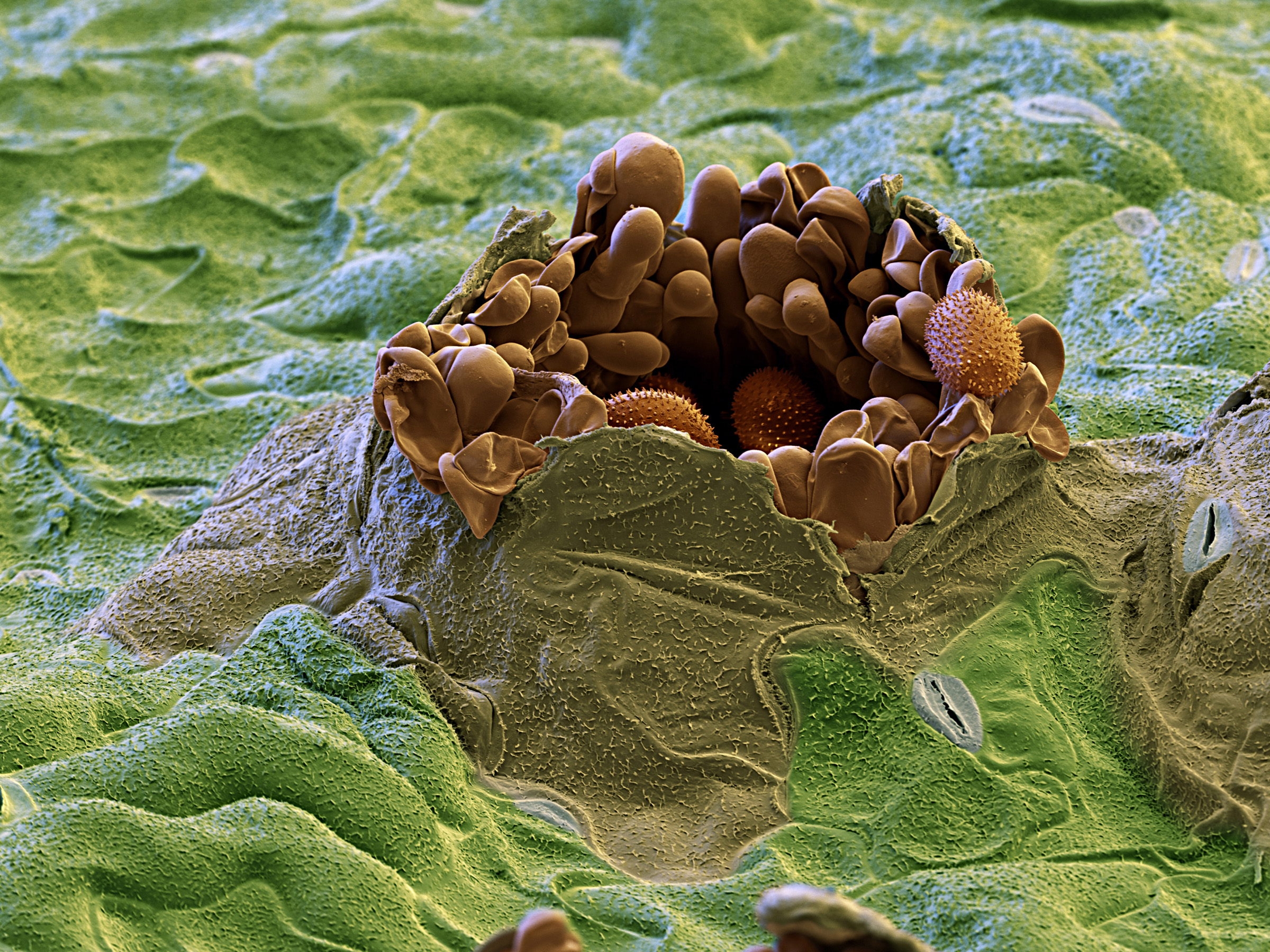
A big problem in Agriculture are rust fungi. This micro-photograph shows rust fungus in a soybean leaf at 1000times magnification.
Rostpilze sind ein grosses Problem in der Landwirtschaft. Mikrofotografie eines Rostpilzes in einem Sojablatt bei 1000facher Vergrösserung.
Methaldehyde

This image made with the light microscope shows the crystals of Methaldehyde. In Farming metaldehyde is used as an agent against slugs.
Diese Aufnahme mit dem Lichtmiktoskop zeigt die Kristalle von Methaldehyd. In der Landwirtschaft wird diese Substanz zur Bekämpfung von Schnecken eingesetzt.
Wheat Germ

Germinating wheat grain shown with the scanning electron microscope. Wheat is grown on more farmland than any other food crop, making it essential to the diet of most of the planet's population.
Keimendes Weizenkorn im Rasterelektronenmikroskop gesehen. Weizen wird auf mehr Ackerfläche angebaut als jede andere Nahrungspflanze und ist somit essentiell für die Ernährung des grössten Teils der Menschheit.
Since we look into the microworld with the eyes of photographers, aesthetics is our highest goal
eye of science can look back on almost three decades of experience in preparation, photography and digitalimaging in scanning electron microscopy. We prepare and photograph samples of chemicals, plants, fungi or plant parasites relevant for harvesting and cultivation using the scanning electron microscope. The goal is the highest possible aesthetic representation while maintaining scientific authenticity. Besides crop and agricultural industry our customers are scientific publishers, chemical and pharmaceutical or the high-tech industry. Via the menu on top you will find examples of our work on electron microscopy, as well as light microscopy and underwater photography, grouped by topic.
eye of science blickt auf fast drei Jahrzehnte Erfahrung in der Präparation, Fotografie und digitaler Bildbearbeitung in der Raster-Elektronenmikroskopie zurück. Wir präparieren und fotografieren mit dem Rasterelektronenmikroskop Proben von Chemikalien, Pflanzen, Pilzen oder Pflanzenparasiten, die für die Ernte und den Anbau relevant sind. Ziel ist eine möglichst hohe ästhetische Darstellung unter Wahrung der wissenschaftlichen Authentizität. Zu unseren Kunden zählen neben der Pflanzen- und Agrarindustrie auch wissenschaftliche Verlage, die Chemie- und Pharmaindustrie sowie die High-Tech-Industrie. Über das Menu oben finden Sie nach Themen gruppiert Beispiele unserer Arbeit am Elektronenmikroskop sowie auch Lichtmikroskopie und Unterwasserfotografie.
© Copyright for all images on this site by eye of science. All rights reserved.